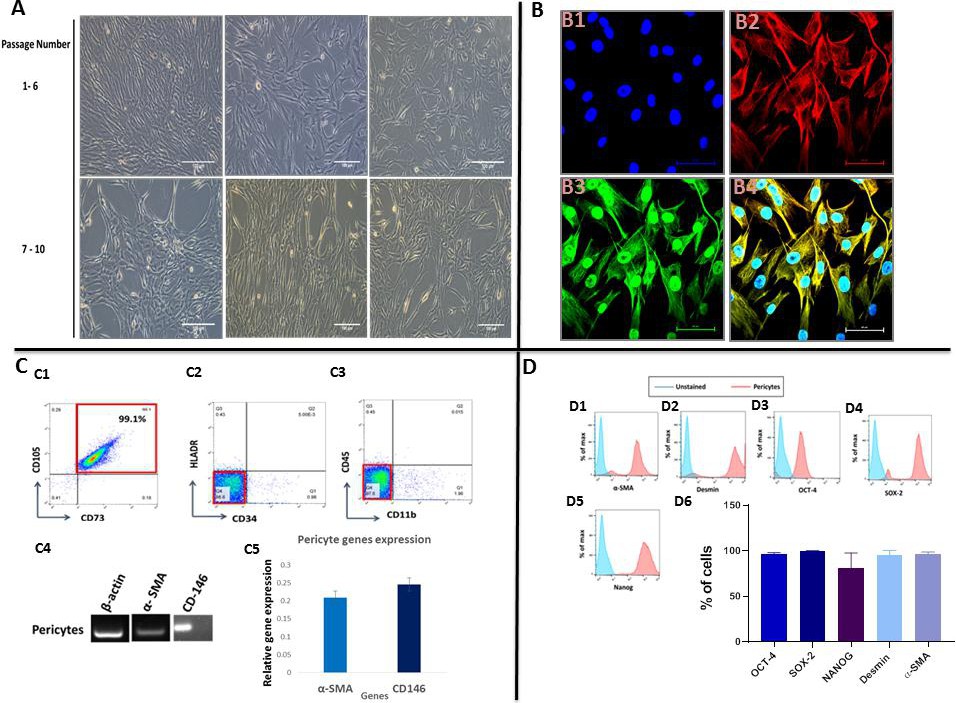
Fig. 2. Morphological, phenotypic and genotypic analyses of pericytes. A. Images of human adipose-derived pericytes at different passages captured using an inverted microscope: (A-C) p1-p6, and (D-F) p7-p10. B. Confocal microscope images showing the expression of alpha smooth muscle actin and desmin. B1. nuclear staining using Hoechst, B2. alpha smooth muscle actin expression, B3. desmin expression, and B4. merged image from B1-B3. C. Flow cytometry analysis of human adipose tissue-derived pericytes. (C1-C3) showing expression of the mesenchymal stem cells markers CD105 and CD73, but not HLA-DR, or the hematopoietic stem cells markers CD34, CD45 and CD11b. C4,5. Conventional PCR analysis showing the expression of alpha smooth muscle actin and CD146. D. Flow cytometry analysis of human adipose tissue-derived pericytes. (D1-D6) showing expression of the pericyte markers desmin and alpha smooth muscle actin and the pluripotency markers Oct4, Sox2 and Nanog.